In this blog post I will am writing on how to make the human respiratory system working model 3D for school science project using syringe available at your home diy – lungs working
#respiratorysystem #workingmodel #lungsmodel #lungsworkingmodel #schoolproject #scienceproject #scienceexhibition #sciencefair #syringe #diy

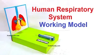
Creating a 3D working model of the human respiratory system using cardboard, syringes, tubing, and balloons can be an engaging and educational project.
Here’s a step-by-step guide on how to make this model:
Materials Needed
- Cardboard (for the base and structure)
- Syringes (2 large ones)
- Flexible plastic tubing
- Balloons (2 for the lungs, 1 for the diaphragm)
- Scissors
- Glue or tape
- Ruler
- Markers or pens
- Plastic bottle (optional, for the chest cavity)
- Rubber bands
- Small clamps or clips (optional, to control airflow)
Step by Step On Making of Lungs Working Model Using Syringe
1. Prepare the Base and Structure
- Base Board:
- Cut a large rectangular piece of cardboard to serve as the base. This will hold the entire setup.
- Chest Cavity:
- Optionally, cut a plastic bottle in half to represent the chest cavity. This can provide a transparent view of the internal workings.
- Alternatively, create a chest cavity using cardboard by forming a box-like structure.
2. Create the Lung and Diaphragm Mechanism
- Lungs:
- Inflate two balloons slightly to represent the lungs.
- Attach the open ends of the balloons to two pieces of flexible plastic tubing. Secure the connection with rubber bands to prevent air from escaping.
- Insert the other end of the tubes into the nozzles of the syringes. Secure them with tape or glue.
- Diaphragm:
- Cut a larger balloon in half and use the bottom part to represent the diaphragm.
- Stretch the diaphragm balloon over the open end of the plastic bottle or the bottom of your cardboard chest cavity.
- Secure it tightly with a rubber band.
3. Assemble the Model
- Chest Cavity and Lungs:
- Place the balloons (lungs) inside the chest cavity (plastic bottle or cardboard structure).
- Ensure the tubing connected to the balloons extends outside the chest cavity for easy access to the syringes.
- Connecting the Syringes:
- Attach the syringes to the ends of the tubes extending from the balloons.
- Make sure the syringes are easily operable by positioning them outside the chest cavity.
- Base Attachment:
- Secure the chest cavity and the syringes onto the cardboard base using glue or tape.
- Ensure the structure is stable and the syringes can be easily pushed and pulled.
4. Demonstrating the Respiratory Process
- Simulating Breathing:
- Push and pull the plungers of the syringes to inflate and deflate the balloons (lungs), simulating inhalation and exhalation.
- When you pull the syringe plunger, the balloon (lung) inflates, representing inhalation.
- When you push the syringe plunger, the balloon deflates, representing exhalation.
- Diaphragm Movement:
- Demonstrate the diaphragm’s role by pressing down on the stretched balloon (diaphragm) at the bottom of the chest cavity.
- Show how the diaphragm’s movement aids in the inflation and deflation of the lungs.
By following these steps, you can create a detailed and interactive 3D working model of the human respiratory system.