
Introduction
An X-ray machine is an important medical imaging device used by doctors to see structures inside the human body. It uses a controlled beam of X-rays to create an image of internal structures, especially bones. X-rays pass through the body, but different tissues absorb different amounts of radiation.
Bones absorb more X-rays and therefore appear brighter on an X-ray image, while softer tissues appear darker or less visible. This working model demonstrates the basic structure and principle of an X-ray machine in a simple and safe way.
Main Concept of the Model
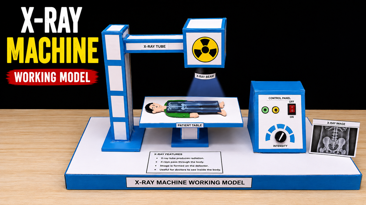
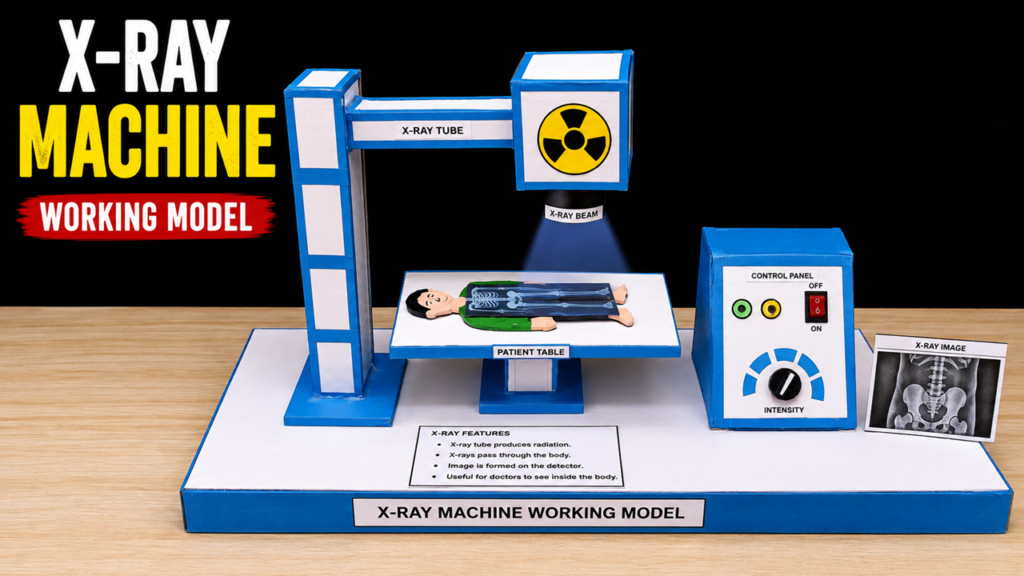
The model consists of an X-ray tube, patient table, control panel, X-ray beam, and detector/image area. The X-ray tube is represented by the large box mounted above the patient table. In a real machine, the X-ray tube produces X-rays when high-speed electrons strike a metal target. The X-rays are directed toward the body.
The patient lies on the table beneath the X-ray tube. In the model, a small human figure with a skeleton image represents the patient. When X-rays pass through the body, different tissues absorb different amounts of radiation. A detector placed behind or below the body receives the X-rays and converts the information into an image. The resulting image can then be viewed by medical professionals.
The control panel represents the part of the machine used by trained radiographers to control settings such as exposure and intensity. The model also includes an X-ray image to show the final result produced by the imaging process.
Materials Required
You can make this project using a large cardboard or foam-board sheet, cardboard boxes, thermocol or foam sheets, white and blue chart paper, glue, scissors, a cutter, ruler, pencil, paints, markers, wooden sticks, plastic tubes, small LEDs, wires, a battery, and printed pictures. A small toy figure or handmade cardboard patient can be used for the patient. Use lightweight materials so the overhead structure remains stable.
How to Make the Model
First, prepare a large rectangular cardboard base. Cover it with white paper and use blue paper around the edges. This will become the main platform of the model.
Next, construct the vertical support column on one side using stacked cardboard pieces. Make sure it is wide and strong enough to support the upper arm. Attach a horizontal cardboard arm extending over the patient table.
At the end of this arm, make a rectangular box representing the X-ray tube. Cover it with white and blue paper and place a radiation warning symbol on the front. Add a label saying “X-RAY TUBE.” A small paper or LED beam can be positioned below it to represent the X-ray beam.
Build the patient table using thick cardboard. Make a rectangular tabletop and support it with a strong central stand. Place a small human figure on the table and add a printed or hand-drawn skeleton image over the figure to represent the body being examined.
On the opposite side, make a separate cardboard box for the control panel. Add paper buttons, an ON/OFF switch, and a circular dial labeled “INTENSITY.” These should only be decorative parts of the school model.
Finally, place a printed X-ray image beside the table and label it “X-RAY IMAGE.” Add an information card explaining the basic steps: X-ray tube produces X-rays → X-rays pass through the body → tissues absorb different amounts → detector receives X-rays → image is produced.
Conclusion
This model explains the basic working principle of an X-ray machine and shows how X-rays help doctors examine structures inside the body. It demonstrates the relationship between the X-ray tube, patient, detector, and control system. The project is useful for understanding medical physics and the role of technology in healthcare.
Safety Note: This is only a demonstration model. Never use a real X-ray tube, radioactive material, or high-voltage X-ray equipment for a school project. LEDs, paper beams, and printed images are sufficient to demonstrate the concept safely.