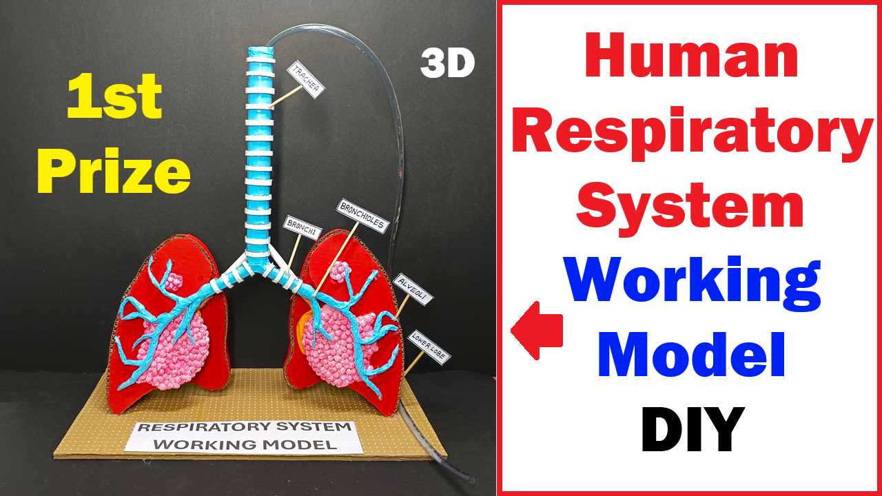
This 3D Human Respiratory System Working Model created for a science project exhibition. The model clearly demonstrates how the human lungs function during breathing. It is designed using simple craft materials like cardboard, colored paper, tubes, and labels, but it effectively explains a very important biological process — respiration.
Let us understand the structure and working of this model in detail.
Introduction to the Human Respiratory System
The human respiratory system is responsible for breathing and gas exchange. It allows us to take in oxygen from the air and remove carbon dioxide from the body. Oxygen is essential for producing energy in our cells, and without it, survival is not possible.
This working model represents the main organs involved in respiration, including:
- Trachea
- Bronchi
- Bronchioles
- Alveoli
- Lungs
Each part is carefully labeled in the model, making it easy to understand the pathway of air movement.
Structure of the Model
The model is built on a rectangular cardboard base labeled “Respiratory System Working Model.” Two red lung-shaped structures are placed on either side. These represent the right and left lungs.
At the top center, there is a vertical tube labeled “Trachea.” This tube splits into two branches labeled “Bronchi.” These bronchi further divide into smaller branches called “Bronchioles.” At the ends of these tiny branches are pink clustered structures labeled “Alveoli.”
A transparent pipe is attached to demonstrate airflow. This pipe helps simulate inhalation and exhalation in the working demonstration.
The neat labeling and color contrast make the model visually attractive and scientifically clear.
Parts of the Respiratory System Explained
1. Trachea (Windpipe)
The trachea is the main air passage that connects the nose and mouth to the lungs. In the model, the vertical blue and white striped tube represents the trachea.
In real life:
- It is made of cartilage rings.
- It prevents the airway from collapsing.
- It allows air to pass into the lungs.
In the model, the trachea acts as the entry point for air.
2. Bronchi
The trachea divides into two main branches called bronchi — one for each lung.
In the model:
- The bronchi are shown as two branching tubes extending into each lung.
- They are labeled clearly.
In real lungs:
- The right bronchus goes to the right lung.
- The left bronchus goes to the left lung.
These bronchi carry air deeper into the lungs.
3. Bronchioles
The bronchi further divide into smaller tubes called bronchioles.
In the model:
- Thin blue branching structures inside the lungs represent bronchioles.
- They look like tree branches spreading throughout the lung.
In real life:
- Bronchioles distribute air evenly inside the lungs.
- They control airflow by expanding or narrowing.
4. Alveoli
At the end of bronchioles are tiny air sacs called alveoli.
In the model:
- The pink clustered sections inside each lung represent alveoli.
- They appear like bunches of grapes.
In real lungs:
- Millions of alveoli are present.
- They have thin walls.
- They are surrounded by blood capillaries.
Alveoli are the most important part of the respiratory system because gas exchange happens here.
Working of the Model
This model demonstrates the process of breathing, which has two main stages:
- Inhalation (Breathing In)
- Exhalation (Breathing Out)
Let us understand both processes.
Inhalation (Breathing In)
During inhalation:
- Air enters through the nose or mouth.
- It passes through the trachea.
- Air moves into the bronchi.
- It travels through bronchioles.
- Finally, it reaches the alveoli.
In real life:
- The diaphragm contracts and moves downward.
- The chest cavity expands.
- Lung volume increases.
- Air pressure inside lungs decreases.
- Air flows inside.
In the working model:
- When air is pushed through the pipe, it flows down the trachea.
- It spreads into both lungs.
- This demonstrates how oxygen enters the respiratory system.
Gas Exchange in Alveoli
Inside the alveoli:
- Oxygen moves from the air into the blood.
- Carbon dioxide moves from the blood into the alveoli.
This happens by diffusion because:
- Oxygen concentration is higher in alveoli.
- Oxygen concentration is lower in blood.
So oxygen naturally moves into the bloodstream.
At the same time:
- Carbon dioxide concentration is higher in blood.
- Carbon dioxide concentration is lower in alveoli.
So carbon dioxide moves into alveoli to be exhaled.
The model visually represents alveoli clusters to explain this important exchange process.
Exhalation (Breathing Out)
During exhalation:
- The diaphragm relaxes and moves upward.
- The chest cavity size decreases.
- Lung volume reduces.
- Air pressure increases inside lungs.
- Carbon dioxide is pushed out.
In the model:
- When air is released from the system, it flows back through bronchioles.
- It passes through bronchi.
- It exits via the trachea.
This shows how carbon dioxide leaves the body.
Importance of the Respiratory System
The respiratory system is essential because:
- It supplies oxygen for cellular respiration.
- It removes carbon dioxide waste.
- It helps maintain blood pH balance.
- It supports speech production.
- It helps regulate body temperature.
Without proper breathing, organs cannot function.
Conclusion
The 3D Human Respiratory System Working Model is an excellent educational project that demonstrates the structure and function of the lungs. It clearly explains the pathway of air from the trachea to alveoli and shows how oxygen enters the body while carbon dioxide leaves.
The model effectively represents inhalation and exhalation using simple craft materials. The labeled parts like trachea, bronchi, bronchioles, and alveoli make the explanation easy to understand.
This project not only enhances scientific knowledge but also improves presentation skills and creativity. If explained confidently with proper understanding of breathing mechanics and gas exchange, this model can definitely win first prize in science exhibitions.
It is a perfect example of how complex biological systems can be demonstrated in a simple, creative, and practical way for learning and teaching purposes.