
Good morning everyone.
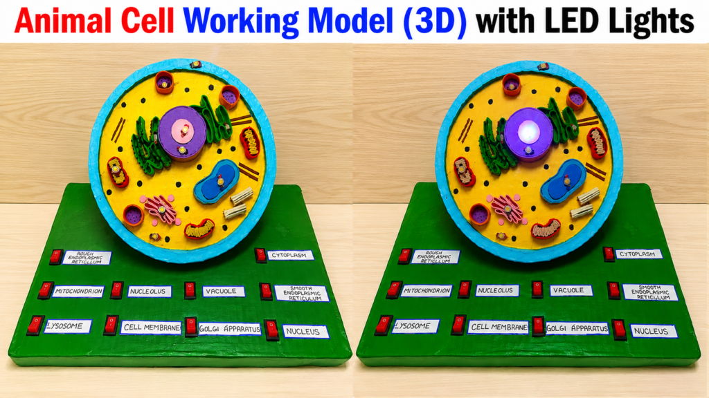
Today I am going to explain my science project on “Animal Cell 3D Working Model with LED Lights.” This DIY model helps students understand the structure and functions of an animal cell in a simple, creative, and interactive way.
A cell is known as the basic structural and functional unit of life. All living organisms are made up of cells. Animal cells are found in animals and human beings. Although cells are very small and can only be seen under a microscope, they perform many important functions necessary for life.
This 3D working model shows the important parts of an animal cell called cell organelles. LED lights are connected to different organelles to make the model attractive and easy to understand during science exhibitions.
The outer blue layer of the model is called the Cell Membrane. It protects the cell and controls the movement of substances in and out of the cell. It acts like a protective covering.
Inside the cell is the yellow-colored Cytoplasm. It is a jelly-like substance where all the organelles are present. Many chemical reactions take place in the cytoplasm.
The large purple structure in the center is the Nucleus. It is called the control center of the cell because it controls all activities of the cell. The nucleus contains genetic material called DNA, which carries hereditary information.
Inside the nucleus is the Nucleolus, which helps in the formation of ribosomes.
The bean-shaped structures shown in red and yellow are the Mitochondria. They are known as the powerhouse of the cell because they produce energy through cellular respiration. This energy helps the cell perform various activities.
The green folded structures represent the Endoplasmic Reticulum (ER). It helps in transporting materials inside the cell. There are two types:
- Rough Endoplasmic Reticulum
- Smooth Endoplasmic Reticulum
The rough ER contains ribosomes and helps in protein synthesis, while the smooth ER helps in fat production.
The pink structure is the Golgi Apparatus. It stores, packages, and transports proteins and other substances to different parts of the cell.
The small round structures shown are Lysosomes and Vacuoles. Lysosomes help in digestion and removal of waste materials, while vacuoles store food, water, and other substances.
The small black dots represent Ribosomes, which help in making proteins for the body.
The LED lights connected to each organelle make the project interactive. When a switch is pressed, the corresponding organelle glows. This helps students identify each part easily and understand its function clearly.
Materials Used
This DIY project is made using:
- Thermocol or foam sheet
- Cardboard
- LED lights
- Battery and wires
- Paint colors
- Glue
Educational Importance
This model helps students learn:
- Cell structure
- Functions of organelles
- Biology concepts
- Electrical circuits using LEDs
- Practical and creative learning
Advantages of the Model
- Easy to understand
- Attractive 3D design
- Interactive learning with lights
- Improves biology knowledge
- Perfect for science exhibitions
Conclusion
In conclusion, this Animal Cell 3D Working Model with LED Lights is an educational and creative science project that explains the structure and functions of an animal cell clearly. The LED lights make learning more interesting and help students identify organelles easily.
This project combines biology with creativity and electronics, making science learning fun and practical for students.