Introduction
The human heart is a vital organ of the circulatory system. It works continuously like a pump to circulate blood throughout the body. The heart supplies oxygen-rich blood to all organs and removes carbon dioxide and waste products. Understanding the working of the heart is very important in biology.

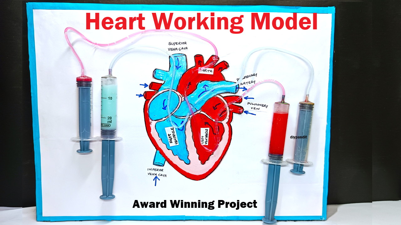
This Human Heart Working Model is a DIY science exhibition project made using cardboard and syringes. The model clearly demonstrates how blood enters the heart, gets pumped, and flows out to different parts of the body. Syringes are used to show blood movement, making the model interactive and easy to understand.
Aim of the Project
The main objectives of this project are:
- To understand the structure and working of the human heart
- To demonstrate blood circulation using a working model
- To explain pumping action of the heart using syringes
- To create a low-cost biology project for science exhibitions
Materials Used
- Cardboard (base and heart structure)
- Color paper or paints (red and blue)
- Three or four syringes (without needles)
- Transparent plastic pipes or IV tubes
- Glue and tape
- Scissors and cutter
- Marker pens for labeling
Structure of the Human Heart
The human heart has four chambers:
- Right atrium
- Right ventricle
- Left atrium
- Left ventricle
The right side of the heart carries impure (deoxygenated) blood, while the left side carries pure (oxygenated) blood.
How to Draw the Heart Picture on Cardboard
- Take a large piece of cardboard
- Draw an outline of the human heart using a pencil
- Divide the heart into four chambers clearly
- Color the right side blue (impure blood)
- Color the left side red (pure blood)
- Draw arrows to show blood flow direction
- Label all chambers and blood vessels neatly
This drawn heart forms the base of the working model.
Description of the Working Model
The heart drawing is mounted vertically on a cardboard base. Syringes are connected using transparent pipes to represent blood vessels.
- Input Syringe (Impure Blood): Represents blood entering the right atrium
- Pump Syringes: Represent ventricular pumping action
- Output Syringe (Pure Blood): Represents blood leaving the left ventricle
Colored water is used to represent blood.
Working Principle of the Model
- Blue-colored water is pushed through the input syringe
- It enters the right side of the heart
- Syringe pumping action represents heart contraction
- Blood moves to the left side after purification (shown conceptually)
- Red-colored water is pushed out through the output syringe
This action demonstrates how the heart pumps blood continuously.
Scientific Principle
The model works on the principle of pressure and pumping action. When a syringe is pressed, pressure is created, pushing the liquid forward, similar to how heart muscles contract to pump blood.
Educational Importance
- Makes heart working easy to understand
- Helps in viva explanations
- Encourages hands-on learning
- Ideal for biology exhibitions
Advantages of Using Syringes
- Clear demonstration of blood flow
- Interactive and engaging
- Low-cost and reusable
Health Awareness
The model helps spread awareness about heart health, importance of exercise, balanced diet, and avoiding unhealthy habits.
Conclusion
The Human Heart Working Model Using Syringes is a simple yet effective science exhibition project. By combining a drawn heart diagram with syringe-based pumping, the model clearly explains the structure and function of the human heart. This DIY project is economical, educational, and perfect for school science exhibitions.
A Healthy Heart Means a Healthy Life.