Introduction
The human digestive system is a group of organs that helps the body break down food, absorb nutrients, and remove waste. It starts from the mouth and ends at the anus.
In the mouth, food is chewed by teeth and mixed with saliva, which contains enzymes to start breaking down starch. The chewed food then passes through the esophagus into the stomach, where strong acids and enzymes digest proteins. The stomach turns the food into a semi-liquid form called chyme.
Next, the food enters the small intestine, where most digestion and absorption take place. Nutrients like vitamins, minerals, proteins, and fats are absorbed into the blood. The remaining undigested food moves into the large intestine, where water is absorbed and waste is formed into feces. Finally, feces are removed from the body through the anus.
Other organs like the liver, pancreas, and gallbladder help by producing bile and enzymes that aid digestion.
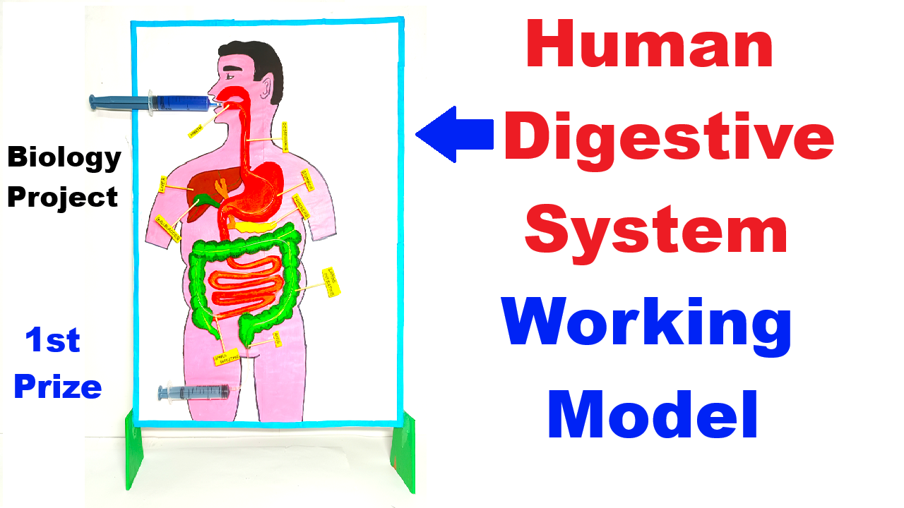
How to Make Human Digestive System
This working model shows how food moves through the human digestive system using peristalsis. Each syringe represents muscle movement that pushes food from one organ to another.
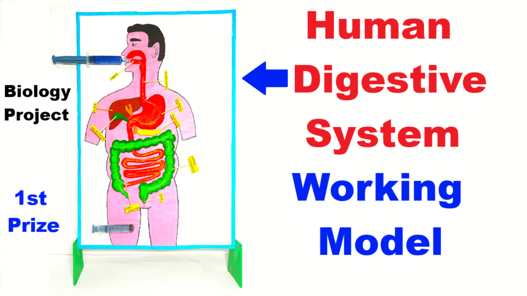
Starting from the mouth, food travels through the esophagus to the stomach, then into the intestines where digestion and absorption take place. Finally, waste is expelled through the rectum.
The model uses cardboard for structure and syringes with tubing to simulate real food movement, helping us understand digestion in a visual and interactive way.
Project Theme:
Demonstrates the journey of food through the digestive system using cardboard organs and syringes to show peristalsis (muscle movement) and digestion.
Materials Required:
- Large cardboard sheet (for base and background)
- Paints (red, pink, brown, green – for organs)
- Syringes (2) (10ml or 20ml)
- IV tubing or aquarium pipes (to connect syringes to simulate food flow)
- Glue / Fevicol / Hot glue
- Small colored beads or thick liquid (like colored water, flour paste, or jelly) – to represent food
- Scissors, marker pens
- Transparent plastic sheet (optional – for stomach/intestinal window)
Video Steps on How to Make the Model (Step-by-Step):
1. Draw and Cut Organ Shapes on Cardboard
- Mouth
- Esophagus
- Stomach
- Small intestine
- Large intestine
- Rectum
Paint them realistically and glue them on a large upright cardboard base in correct anatomical order.
2. Set Up Syringe-Driven Flow Mechanism
- Use tubing and syringes to simulate food movement:
- Connect:
- Syringe → mouth to rectum (for final ejection)
- Paste the syringes on the side or behind the board.
- Ensure tubes are transparent and fixed along the organ pathway.
- Connect:
3. Add the “Food”
- Use a thick liquid (colored flour paste or jelly-like substance) or small colored beads to represent chewed food.
- Insert this from the mouth opening.
4. Working Mechanism (Peristalsis Simulation)
- When you pull and push the syringes in sequence, it will push the food forward just like muscles do in real digestion.
- You can demonstrate:
- Swallowing (mouth to stomach)
- Churning (stomach to intestine)
- Absorption and waste removal