
Good morning everyone.
Today I am going to explain my science project on “Animal Cell 3D DIY Model.” This project helps students understand the structure and functions of an animal cell in a simple and creative way.
A cell is called the basic structural and functional unit of life. All living organisms are made up of cells. Animal cells are found in animals and human beings. They are microscopic in size, but they perform very important functions that keep the body alive.
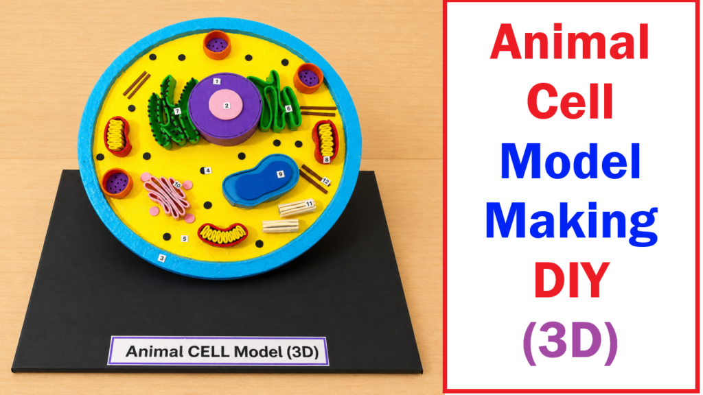
This 3D model shows the internal structure of an animal cell along with its important organelles. Different colors and shapes are used to make the parts easy to identify and understand.
The outer blue boundary is called the Cell Membrane. It protects the cell and controls the movement of substances in and out of the cell. It acts like a security guard for the cell.
The yellow area inside the cell is called the Cytoplasm. It is a jelly-like substance where all cell organelles are present. Various chemical reactions take place in the cytoplasm.
The large purple structure in the center is the Nucleus. The nucleus is known as the control center of the cell because it controls all activities of the cell. It also contains genetic material called DNA, which carries hereditary information from parents to offspring.
Inside the nucleus is a smaller structure called the Nucleolus, which helps in the formation of ribosomes.
The red and yellow bean-shaped structures are called Mitochondria. They are known as the powerhouse of the cell because they produce energy through respiration. This energy helps the cell perform various functions.
The green folded structures represent the Endoplasmic Reticulum (ER). It helps in the transport of materials inside the cell. There are two types of ER:
- Rough ER
- Smooth ER
The rough ER contains ribosomes, while the smooth ER helps in lipid production.
The small black dots in the model are Ribosomes. Ribosomes help in protein synthesis. Proteins are necessary for growth and repair of the body.
The pink stacked structure is the Golgi Apparatus. It stores, modifies, and packages proteins and other substances before sending them to different parts of the cell.
The small circular structures shown in the model are Lysosomes and Vacuoles. Lysosomes help in digestion and removal of waste materials, while vacuoles store food, water, and other substances.
The rod-like structures represent the Centrioles, which help during cell division.
This DIY project is made using simple materials like:
- Foam sheets
- Cardboard
- Clay
- Paint colors
- Glue
This project helps students learn science through practical activity and visual understanding. It improves creativity, observation skills, and knowledge about biology.
Advantages of This Model
- Easy to understand
- Attractive 3D design
- Interactive learning
- Improves biology knowledge
- Useful for school exhibitions
Importance of Animal Cells
Animal cells help in:
- Growth of the body
- Energy production
- Transport of materials
- Repair of tissues
- Reproduction
In conclusion, this Animal Cell 3D DIY Model is an educational and creative project that explains the structure and functions of animal cell organelles clearly. It makes biology learning more interesting and enjoyable for students.