
The digestive system is a complex and vital system in the human body responsible for breaking down food into smaller, absorbable components and extracting nutrients for energy, growth, and repair.
It involves various organs and processes that work together to facilitate the digestion and absorption of food.
Main Organs and Their Functions:
- Mouth: The process of digestion begins in the mouth, where food is broken down mechanically by chewing and mixed with saliva. Saliva contains enzymes like amylase, which starts the chemical breakdown of carbohydrates.
- Esophagus: The esophagus is a muscular tube that connects the mouth to the stomach. It transports chewed and moistened food to the stomach through a series of coordinated muscular contractions called peristalsis.
- Stomach: The stomach is a muscular organ that further breaks down the food through churning and mixing with gastric juices. Gastric juices contain hydrochloric acid and enzymes, such as pepsin, which help break down proteins.
- Small Intestine: The small intestine is the primary site of digestion and nutrient absorption. It consists of three parts: the duodenum, jejunum, and ileum. The pancreas and liver secrete digestive enzymes and bile, respectively, into the small intestine. These enzymes and bile aid in the breakdown of fats, proteins, and carbohydrates into their smaller components (fatty acids, amino acids, and glucose), which are then absorbed into the bloodstream through the intestinal lining.
- Pancreas: The pancreas is both an endocrine and exocrine gland. As an exocrine gland, it produces and secretes digestive enzymes (lipase, protease, and amylase) into the small intestine to aid in digestion.
- Liver: The liver plays a crucial role in digestion by producing bile, which is stored in the gallbladder and released into the small intestine. Bile helps emulsify fats, breaking them into smaller droplets, making it easier for enzymes to digest them.
- Gallbladder: The gallbladder stores bile produced by the liver and releases it into the small intestine when needed for digestion.
- Large Intestine (Colon): The remaining undigested food and waste move into the large intestine, where water and electrolytes are absorbed, and the remaining material is formed into feces. Beneficial bacteria in the large intestine also aid in the fermentation of undigested carbohydrates, producing gases and certain vitamins.
- Rectum and Anus: The rectum stores feces until it is ready to be expelled from the body through the anus during the process of defecation.
The digestive system is a highly coordinated and efficient system that ensures the body receives the necessary nutrients from the food we eat while eliminating waste products. Proper digestion and absorption of nutrients are essential for maintaining good health and well-being.
#digestivesystemworkingmodel #workingmodel #scienceexhibition #scienceproject #sciencemodel #sciencefair
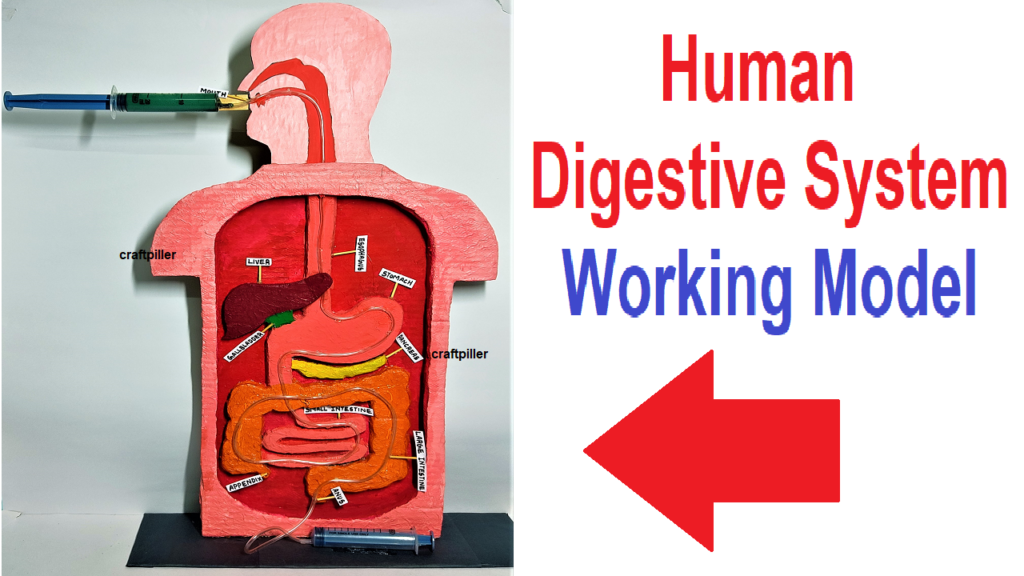
Step by step video on making of digestive system working model
Creating a digestive system model project that helps you understand the various organs and processes involved in digestion.
Here’s a step-by-step guide to making a simple digestive system model:
Materials Needed:
Large cardboard
Colored construction cardboard with paper (to represent organs)
Scissors
Glue
Markers or colored pencils (for labeling)
Syringes to represent food flow
Step-by-Step Instructions:
Prepare the Base: Take a large cardboard sheet as the base for your model. This will serve as the canvas for creating the different parts of the digestive system.
Draw and Cut Out Organs: On colored construction cardboard / paper , draw and cut out the various organs of the digestive system. Here are the essential organs to include:
- Mouth
- Esophagus
- Stomach
- Small intestine (you can cut it into three parts: duodenum, jejunum, and ileum)
- Pancreas
- Liver
- Gallbladder
- Large intestine (colon)
- Rectum and Anus
Arrange the Organs and place the syringes : Position the cut-out organs on the cardboard base in the correct order to represent the digestive system. Arrange them in a line from the mouth to the anus, simulating the pathway that food takes through the body during digestion.
Add Labels: Use markers or colored pencils to label each organ with its name. You can also add arrows or lines to indicate the direction of food flow.