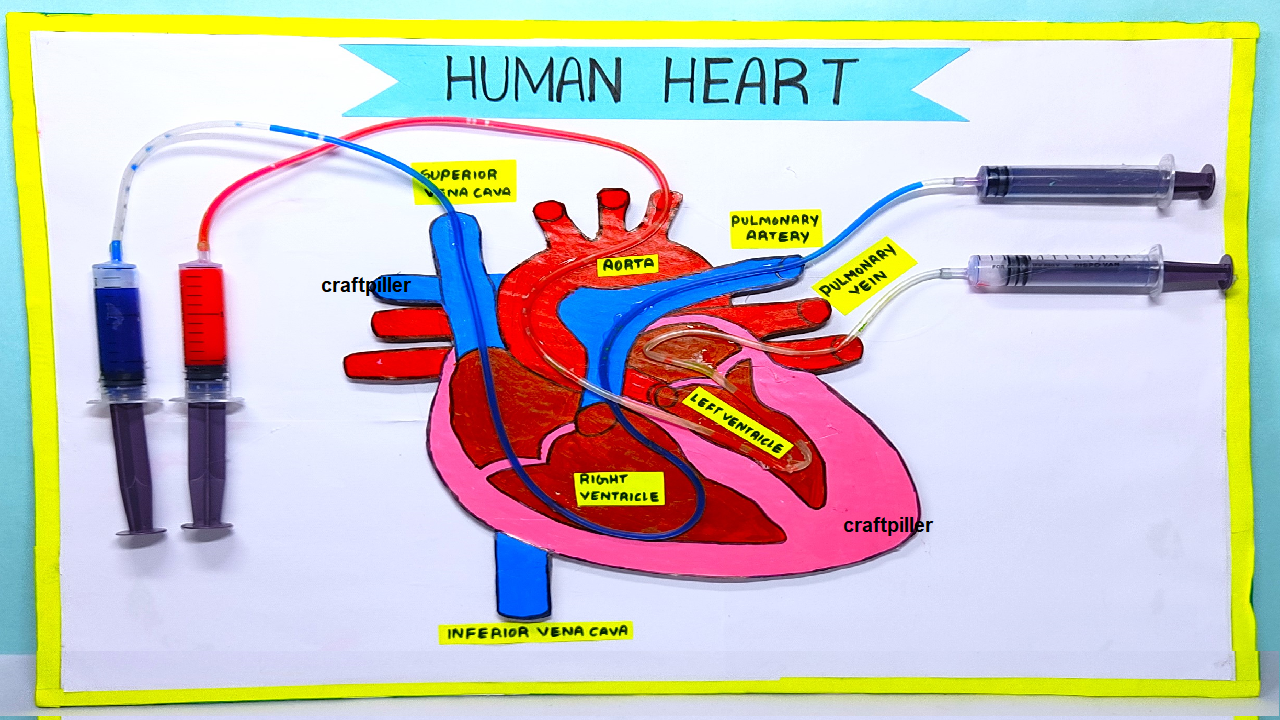
Using syringes to model the human heart is an effective way to demonstrate how the heart functions. Here are some likely questions you might encounter at a science exhibition, along with detailed answers:

1. How does your model represent the human heart?
Answer:
My model uses syringes to represent the chambers of the heart and simulate its pumping action. Each syringe represents a different chamber of the heart:
- Two syringes act as the left and right atria.
- Two syringes act as the left and right ventricles. The syringes are connected by tubes to mimic the flow of blood through the heart, and their plunger movements simulate the heart’s contractions and relaxations.
2. How does the model simulate the heart’s pumping action?
Answer:
The model simulates the heart’s pumping action through the operation of the syringes:
- Filling: When a syringe’s plunger is pulled back, it represents the heart’s relaxation phase (diastole), allowing it to fill with “blood” (liquid).
- Pumping: When the plunger is pushed forward, it represents the heart’s contraction phase (systole), forcing the liquid out into the connected tubes, simulating blood being pumped out of the heart.
3. What do the syringes and tubes represent in terms of heart anatomy?
Answer:
- Syringes: Represent the heart’s chambers. For example, one syringe for the right atrium, another for the right ventricle, and similarly for the left atrium and left ventricle.
- Tubes: Represent the blood vessels. For instance, tubes connecting the syringes simulate the pulmonary artery, pulmonary veins, aorta, and vena cavae.
4. How does your model show the flow of blood through the heart?
Answer:
The model demonstrates the flow of blood through the heart by showing how blood moves from one chamber to the next:
- Deoxygenated Blood Flow: Syringe representing the right atrium fills up and then pumps blood into the right ventricle, which then sends blood through the pulmonary artery to the lungs.
- Oxygenated Blood Flow: Blood returns to the left atrium, which then pumps it into the left ventricle. The left ventricle then pumps the blood into the aorta and throughout the body.
5. How do the valves in the heart work, according to your model?
Answer:
In the model, check valves (one-way valves) could be represented by simple mechanisms or flaps that prevent backflow:
- Tricuspid Valve: Between the right atrium and right ventricle.
- Pulmonary Valve: Between the right ventricle and the pulmonary artery.
- Mitral Valve: Between the left atrium and left ventricle.
- Aortic Valve: Between the left ventricle and the aorta.
These valves ensure that blood flows in only one direction and does not backtrack into the previous chamber.
6. How does the model show the difference between oxygenated and deoxygenated blood?
Answer:
The model can use different colored liquids or dyes to represent oxygenated and deoxygenated blood:
- Blue Liquid: Represents deoxygenated blood returning from the body.
- Red Liquid: Represents oxygenated blood being pumped to the body.
This visual distinction helps to illustrate how blood changes in oxygen content as it travels through the heart and lungs.
7. What are the limitations of your model?
Answer:
While the model effectively demonstrates the basic function of the heart, it has limitations:
- Simplification: It does not fully capture the complex interactions and regulation of the heart’s function.
- Scale: The syringes and tubes do not represent the true scale of the heart and blood vessels.
- Detail: It may not show all the details of blood flow or heart conditions accurately.
8. How does this model help in understanding heart diseases?
Answer:
The model can help visualize how blockages or defects might affect heart function:
- Valve Malfunction: You can show how a valve malfunction might cause backflow or inefficiency.
- Blocked Artery: By demonstrating how blocked tubes (simulating arteries) affect blood flow, you can explain how arterial blockages impact the heart.
9. How can this model be used for educational purposes?
Answer:
This model is useful for teaching:
- Basic Heart Anatomy: It provides a visual and interactive representation of the heart’s chambers and valves.
- Circulatory System Function: It helps to demonstrate how blood circulates through the heart and the body.
- Heart Health: It can be used to discuss the importance of heart health and how lifestyle choices impact heart function.
10. How did you construct the model and what materials did you use?
Answer:
The model was constructed using:
- Syringes: To represent the heart’s chambers.
- Plastic Tubing: To simulate blood vessels.
- Colored Liquids: To differentiate between oxygenated and deoxygenated blood.
- Check Valves or Flaps: To mimic heart valves and prevent backflow.

