The human blood circulatory system, also known as the cardiovascular system, is a complex network of vessels, the heart, and other organs that work together to transport oxygen, nutrients, hormones, and waste products throughout the body.

The circulatory system can be divided into two main components: the systemic circulation and the pulmonary circulation.
1. Systemic Circulation:
a. Function:
- The systemic circulation is responsible for delivering oxygenated blood, rich in nutrients, to the body’s tissues and organs. It also collects deoxygenated blood, containing waste products, and returns it to the heart for oxygenation.
b. Process:
- Oxygenated blood from the left ventricle of the heart is pumped into the main artery, the aorta.
- The aorta branches out into smaller arteries, which further divide into arterioles, delivering oxygenated blood to tissues.
- In capillaries, which are tiny blood vessels, oxygen and nutrients are exchanged for waste products.
- Deoxygenated blood is collected from the tissues and carried back through venules to veins.
- Veins converge into the superior and inferior vena cava, which return the deoxygenated blood to the right atrium of the heart.
2. Pulmonary Circulation:
a. Function:
- Pulmonary circulation is responsible for exchanging carbon dioxide for oxygen in the lungs. It ensures that oxygenated blood is returned to the left side of the heart.
b. Process:
- Deoxygenated blood from the body enters the right atrium of the heart through the superior and inferior vena cava.
- The right atrium contracts, pushing blood into the right ventricle.
- The right ventricle contracts and pumps deoxygenated blood into the pulmonary artery.
- The pulmonary artery carries blood to the lungs, where carbon dioxide is exchanged for oxygen.
- Oxygenated blood returns to the heart through the pulmonary veins, entering the left atrium.
3. The Heart:
a. Structure:
- The heart is a muscular organ divided into four chambers: the right atrium, right ventricle, left atrium, and left ventricle.
- The septum separates the right and left sides of the heart.
b. Function:
- The right side of the heart pumps deoxygenated blood to the lungs for oxygenation (pulmonary circulation).
- The left side of the heart pumps oxygenated blood to the rest of the body (systemic circulation).
4. Blood Vessels:
a. Arteries:
- Arteries carry blood away from the heart.
- The aorta is the largest artery, carrying oxygenated blood to the body.
b. Veins:
- Veins carry blood toward the heart.
- The superior and inferior vena cava bring deoxygenated blood from the body to the right atrium.
c. Capillaries:
- Capillaries are tiny blood vessels where the exchange of oxygen, nutrients, and waste products occurs between blood and tissues.
5. Lungs:
a. Function:
- The lungs are the site of gas exchange, where oxygen is taken in, and carbon dioxide is expelled.
b. Process:
- Deoxygenated blood from the body travels to the lungs through the pulmonary artery.
- In the lungs, oxygen is absorbed, and carbon dioxide is released as waste.
- Oxygenated blood returns to the heart via the pulmonary veins.
The coordinated functioning of the heart, blood vessels, and lungs ensures a continuous and efficient circulation of blood throughout the body, supporting the metabolic needs of cells and maintaining homeostasis.
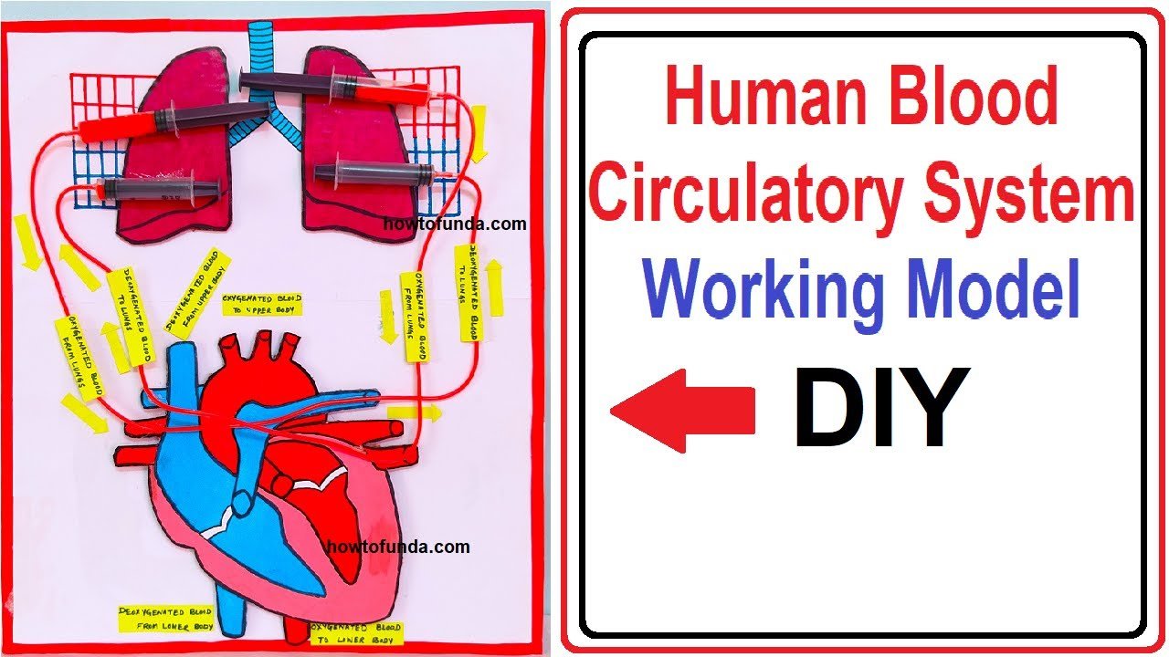
This simple human blood circulatory system working model (heart & lungs) is a great visual aid to help explain the complex workings of the human circulatory system
By creating a working model of the human circulatory system can be a great way to understand for students on how the heart, blood vessels, and blood work together to transport oxygen and nutrients throughout the body.
Here are the video steps to create a simple working model of the human blood circulatory system:
#bloodcirculatorysystem #bloodcirculation #heart #circulatorysystem #heart #lungs #biologyproject #biologymodel #scienceproject #exhibition #diy
Materials Required
Creating a working model of the human blood circulatory system, including the heart and lungs, can be an engaging and educational project. Here’s a simplified version using common materials:
- Large cardboard or poster board
- Red and blue markers or colored paper
- Craft supplies (scissors, glue, tape)
- Small balloons (representing the lungs)
- Plastic tubing or straws
- Small plastic cups
- Water
- Small rubber bands
- Marker pen
- Small toy heart (optional)
Steps:
1. Prepare the Base:
- Use a large piece of cardboard or poster board as the base for your model. This will represent the body.
2. Create the Heart:
- Draw or cut out a simple representation of the heart. You can draw two connected circles, representing the left and right sides of the heart. Alternatively, you can use a small toy heart if available.
3. Color the Blood Vessels:
- Use red markers or colored paper to create blood vessels on the cardboard. Draw arteries leaving the heart and branching into smaller vessels (representing the systemic circulation). Draw veins returning to the heart (representing the pulmonary circulation).
4. Attach Balloon Lungs:
- Blow up two small balloons to represent the lungs. Attach the balloons to the cardboard with tape, placing them on either side of the heart.
5. Connect the Lungs:
- Use plastic tubing or straws to connect the balloons to the heart. Attach one end to the balloons and the other end to the heart, indicating the path of oxygenated and deoxygenated blood.
6. Add Blood Flow Arrows:
- Draw arrows on the blood vessels to indicate the direction of blood flow. Use red for oxygenated blood leaving the heart and blue for deoxygenated blood returning to the heart.
7. Create Capillaries:
- Draw or cut out small capillaries using red markers or colored paper. Attach them between the arteries and veins to represent the capillary network where gas exchange occurs.
8. Add Plastic Cups for Blood Collection:
- Attach small plastic cups to the veins returning to the heart. These cups will collect the “deoxygenated blood” (water).
9. Demonstrate Blood Circulation:
- Pour water into the plastic cups to represent deoxygenated blood returning to the heart. Squeeze the balloons (lungs) to simulate breathing and demonstrate the flow of blood between the heart and lungs.
10. Explain the Model:
- Use a marker pen to label the different parts of the circulatory system, including the heart, lungs, arteries, veins, and capillaries. Explain the functions of each part as you demonstrate the model.
This working model provides a visual representation of the human blood circulatory system, emphasizing the flow of blood between the heart and lungs. It’s a hands-on way to understand the basic concepts of systemic and pulmonary circulation.