Introduction
The human digestive system is one of the most important systems of the body. It helps in breaking down the food we eat into simpler substances so that our body can absorb nutrients and use them for energy, growth, and repair. Without digestion, our body would not be able to get the required nutrients from food.
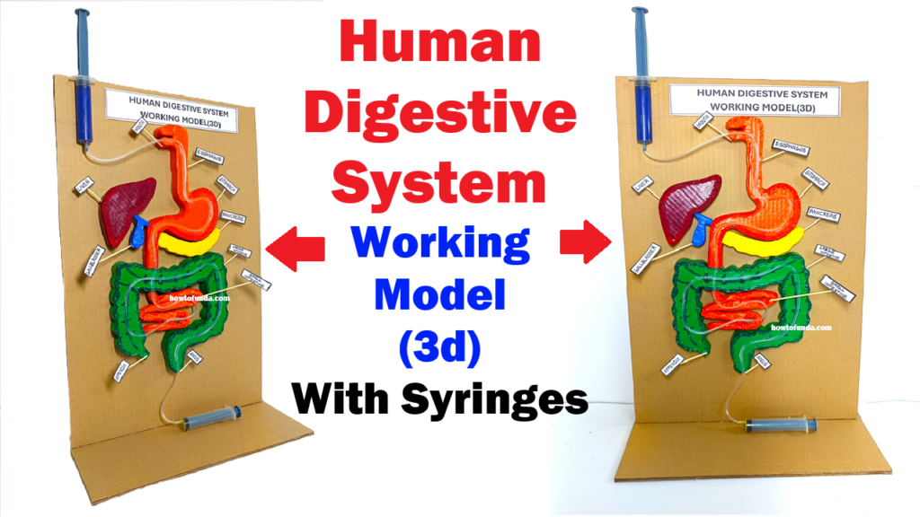
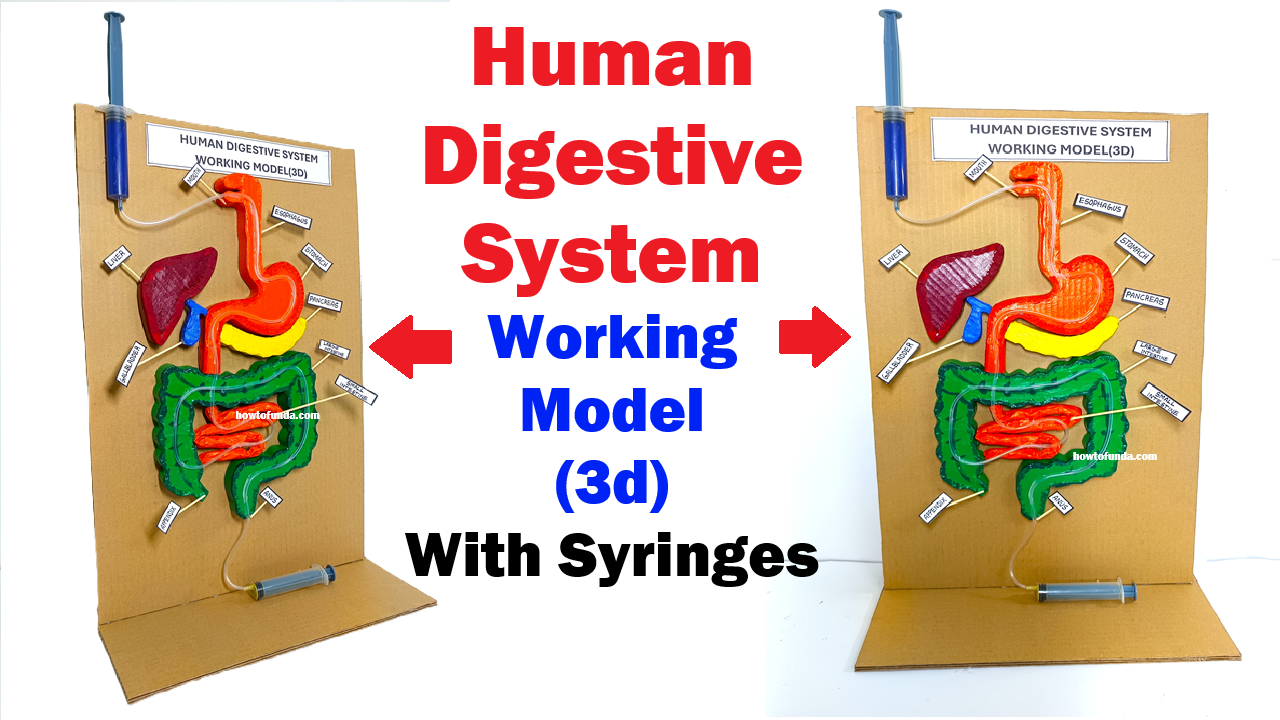
To understand this process in a simple and practical way, this project presents a 3D working model of the human digestive system using syringes, pipes, balloons, and cardboard. This model demonstrates how food moves from the mouth to the stomach and intestines using a mechanism similar to peristalsis (the wave-like movement of muscles in the food pipe). The working model makes learning more interesting because instead of just seeing a diagram, we can actually see the movement of food through the digestive tract.
This project is suitable for school science exhibitions, biology practical demonstrations, CBSE/ICSE projects, and science fairs. It is low-cost, easy to make, and clearly explains the working of the digestive system.
Aim of the Project
The aim of this project is:
- To create a 3D working model of the human digestive system.
- To demonstrate how food travels through different parts of the digestive system.
- To explain the process of digestion, peristalsis, absorption, and waste removal using a simple syringe-based mechanism.
- To make learning biology interactive, visual, and easy to understand.
Principle Behind the Model
The human digestive system works by:
- Ingestion – Taking food into the mouth
- Digestion – Breaking food into simpler substances
- Absorption – Absorbing nutrients into the blood
- Egestion – Removing waste from the body
In our body, the food pipe (esophagus) and intestines move food forward using peristaltic movement. This is a wave-like contraction and relaxation of muscles.
In this model, syringes and flexible pipes are used to:
- Push and pull air or liquid
- Create pressure
- Move colored liquid (which represents food) through the system
This simulates how muscles push food through the digestive tract in real life.
Materials Required
- Cardboard or thermocol sheet (for base and body structure)
- 3 to 5 large syringes (without needles)
- Transparent plastic pipes or IV tubes
- Small balloons or rubber tubes
- Fevicol or hot glue
- Cutter and scissors
- Color paper or paints
- Sketch pens or markers
- Tape
- Colored water (to represent food)
- Chart paper for labeling parts
Parts of the Digestive System Shown in the Model
This model shows the following main parts:
- Mouth
- Esophagus (food pipe)
- Stomach
- Small intestine
- Large intestine
- Rectum and anus
Each part is either represented using cardboard shapes or pipes and balloons to show the path of food.
Construction of the Model
- Base and Structure
First, take a large cardboard sheet and make a standing support or human body outline. Draw or paste a cut-out shape of the human torso. This will be the base on which all digestive organs are fixed. - Mouth and Esophagus
At the top, make a small funnel or opening to represent the mouth. Attach a transparent pipe or tube from the mouth going downward. This pipe represents the esophagus. - Stomach
Use a balloon or a small plastic bottle to represent the stomach. Fix it in the correct position on the cardboard. Connect the esophagus pipe to this stomach balloon. - Small Intestine
Take a long transparent pipe and coil it in a zig-zag or circular shape to represent the small intestine. Fix it properly on the board. - Large Intestine
Use a slightly thicker pipe or make a boundary around the small intestine path to show the large intestine. Connect it to the end of the small intestine. - Syringe Mechanism
Attach syringes at different points (for example near the esophagus and stomach or intestines). These syringes will be used to push air or colored water to move the “food” through the pipes. - Finishing and Labeling
Paint the model neatly and label all parts: Mouth, Esophagus, Stomach, Small Intestine, Large Intestine, Rectum, etc.
Working of the Model
- Ingestion (Mouth)
Colored water is poured into the mouth opening. This colored water represents the food we eat. - Movement through Esophagus
By gently pushing the syringe, pressure is created inside the pipe. This pushes the colored water downward, showing how food moves through the esophagus by peristalsis. - In the Stomach
The colored water enters the balloon or bottle that represents the stomach. Here, we can explain that in real life, the stomach mixes food with digestive juices and breaks it down into simpler substances. - Small Intestine
When we press the next syringe, the liquid moves into the long coiled pipe (small intestine). This shows how digested food slowly moves forward. In the real body, most nutrients are absorbed here into the blood. - Large Intestine
Finally, the liquid reaches the large intestine section. Here we explain that water is absorbed and waste material is formed. - Egestion
The remaining liquid or waste moves towards the end, showing how waste is removed from the body.
Thus, by using syringes, we can clearly demonstrate the step-by-step movement of food through the digestive system.
Explanation of Digestive Process (Biology Concept)
- Mouth: Food is chewed and mixed with saliva. Saliva contains enzymes that start digestion.
- Esophagus: Food moves down by peristaltic movement.
- Stomach: Food is mixed with gastric juices and broken down into a semi-liquid form.
- Small Intestine: Digestion is completed and nutrients are absorbed into the blood.
- Large Intestine: Water is absorbed and waste is formed.
- Rectum and Anus: Waste is removed from the body.
This model helps students understand these steps in a visual and practical way.
Advantages of This Model
- Low-cost and easy to make
- Reusable for demonstrations
- Makes learning fun and interactive
- Clearly shows the movement of food
- Perfect for science exhibitions and fairs
- Helps in better understanding than charts or diagrams
Conclusion
The 3D Human Digestive System Working Model using Syringes is a simple, effective, and educational project that explains one of the most important systems of the human body. By using syringes and pipes, we can clearly demonstrate how food moves through the digestive tract and how digestion takes place step by step.
This model not only improves understanding of biology concepts like peristalsis, digestion, and absorption, but also encourages students to learn through hands-on activities. It proves that science can be learned in a fun, creative, and practical way using simple materials.