
Introduction
Cells are the basic structural and functional units of all living organisms. Every living being, including animals and humans, is made up of millions of tiny cells that perform different biological functions necessary for life. Understanding the structure of a cell is an important part of biology education, and creating a 3D animal cell model is an effective way to visualize and learn about the different parts of a cell.
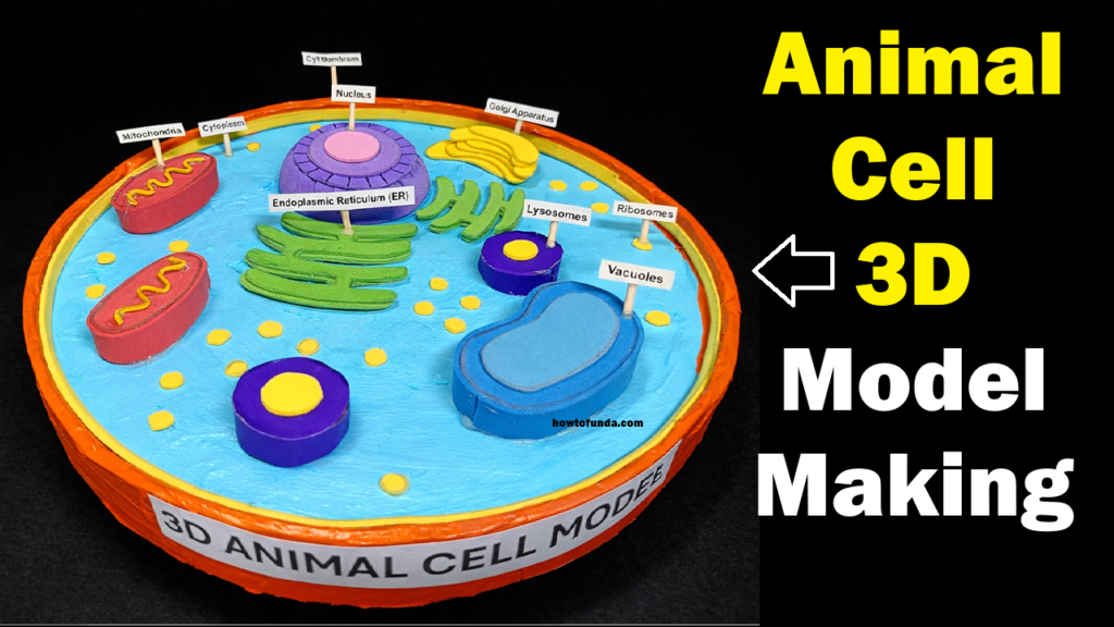
The model shown in the image represents a three-dimensional animal cell, designed for a science exhibition or biology project. This model displays various cell organelles such as the nucleus, mitochondria, endoplasmic reticulum, Golgi apparatus, ribosomes, lysosomes, vacuoles, cytoplasm, and cell membrane. Each component is labeled clearly so that students can easily understand the structure and functions of the animal cell.
This project helps students learn biology concepts in a creative and practical way.
Structure of the Animal Cell
An animal cell contains several specialized structures called organelles, each performing a specific function. The 3D model demonstrates the following important components.
Cell Membrane
The outer boundary of the cell is called the cell membrane. It protects the cell and controls the movement of substances in and out of the cell.
Cytoplasm
The cytoplasm is the jelly-like substance that fills the inside of the cell. All the organelles are suspended in the cytoplasm, and many cellular activities occur here.
Nucleus
The nucleus is the control center of the cell. It contains genetic material (DNA) that regulates cell growth, metabolism, and reproduction.
Mitochondria
Mitochondria are known as the powerhouses of the cell. They produce energy required for cellular processes by converting nutrients into usable energy.
Endoplasmic Reticulum (ER)
The endoplasmic reticulum is a network of membranes that helps in the transport of proteins and other substances inside the cell.
Golgi Apparatus
The Golgi apparatus modifies, packages, and distributes proteins and lipids to different parts of the cell.
Ribosomes
Ribosomes are tiny structures responsible for protein synthesis. They help produce proteins that are essential for cell growth and repair.
Lysosomes
Lysosomes contain digestive enzymes that break down waste materials and damaged cell parts.
Vacuoles
Vacuoles are storage sacs that store water, nutrients, and waste products inside the cell.
Materials Required
To make this animal cell model, the following materials can be used:
- Thermocol or cardboard base
- Colored foam sheets or clay
- Acrylic paints and brushes
- Glue gun or strong adhesive
- Cutter and scissors
- Small label sticks or toothpicks
- Chart paper for labels
These materials help create a colorful and visually appealing model that clearly represents each cell organelle.
Steps to Make the Model
Step 1: Prepare the Base
Start by taking a circular thermocol or cardboard base. Paint the base with light blue color to represent the cytoplasm. This will form the main body of the cell.
Step 2: Create the Cell Membrane
Build a circular boundary around the edge of the base using foam sheets or clay. Paint it with a bright color such as orange to represent the cell membrane.
Step 3: Make the Nucleus
Create the nucleus using clay or foam sheets. Place it near the center of the cell and paint it with a contrasting color like purple. Add a smaller circle inside it to represent the nucleolus.
Step 4: Add Organelles
Use colored foam or clay to create different organelles:
- Oval shapes for mitochondria
- Folded structures for endoplasmic reticulum
- Stacked curved shapes for Golgi apparatus
- Small dots for ribosomes
- Small round shapes for lysosomes
- Larger sacs for vacuoles
Arrange these organelles carefully inside the cytoplasm.
Step 5: Label the Parts
Attach small label sticks with the names of each organelle. Labeling helps viewers easily identify each part of the animal cell.
Step 6: Final Decoration
Ensure that all parts are neatly arranged and clearly visible. Add bright colors to make the model attractive and easy to understand.
Educational Importance of the Model
This 3D animal cell model helps students visually understand the complex structure of a cell. Instead of learning only from textbooks, students can observe how different organelles are arranged and how they function together.
Such models improve:
- conceptual understanding
- creativity
- scientific presentation skills
- interest in biology
They also help teachers explain difficult biological concepts in a simple way.
Conclusion
The 3D animal cell model is an excellent educational project for science exhibitions and biology learning. It clearly demonstrates the structure and functions of various cell organelles in a visual and interactive way. By building this model, students gain a better understanding of how cells function as the basic units of life.
Hands-on science projects like this encourage curiosity, creativity, and deeper learning. They also help students develop practical skills while exploring important scientific concepts. Understanding the structure of cells is essential for studying biology, medicine, and life sciences, making this project both educational and engaging.