Introduction
Teeth play a vital role in our daily life by helping us chew food, speak clearly, and maintain facial structure. Understanding the internal structure of a tooth is important in biology and health education.
The Tooth Structure 3D Model is a creative and informative project that visually represents the different layers and parts of a human tooth. This model is ideal for school science exhibitions, making it easier for students to understand complex dental anatomy in a simple and engaging way.

Materials Required
This model can be prepared using basic craft materials:
- Cardboard or thermocol base
- Clay or foam sheets
- Acrylic paints or color paper
- Toothpicks (for labeling)
- Glue and scissors
- Marker for writing labels
Working Principle / Concept
The model is based on the anatomy of a human tooth, showing its internal and external structure. Each layer of the tooth has a specific function, and the model highlights these layers using different colors and labels.
The main parts of the tooth included in the model are:
- Enamel
- Dentin
- Pulp cavity
- Root canal
- Gums
- Cementum
- Nerves and blood vessels
This model helps demonstrate how each part works together to maintain the health and function of the tooth.
Structure of the Tooth
- Enamel:
The outermost layer of the tooth and the hardest substance in the human body. It protects the inner layers from damage and decay. - Dentin:
Located beneath the enamel, dentin is softer and supports the enamel. It carries sensations such as pain and temperature. - Pulp Cavity:
The innermost part of the tooth that contains nerves and blood vessels. It is responsible for supplying nutrients and sensation. - Root Canal:
A passage that extends from the pulp cavity to the roots, carrying nerves and blood vessels. - Gums (Gingiva):
Soft tissue that surrounds and protects the base of the teeth. - Cementum:
A layer covering the root of the tooth, helping to anchor it firmly in the jawbone. - Bone and Nerves:
These support the tooth and provide nutrients and sensitivity.
Model Explanation
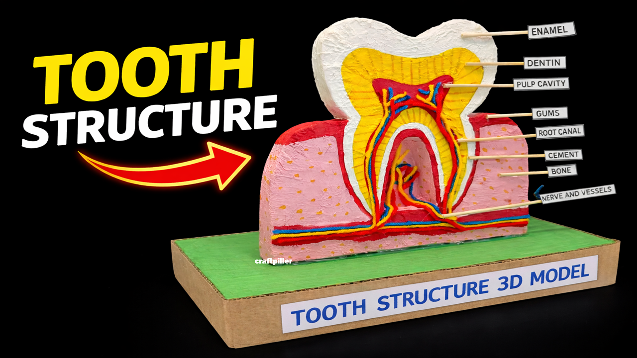
The 3D model shows a cross-sectional view of a tooth mounted on a base. Each layer is clearly represented using different colors:
- The outer white layer represents enamel.
- The yellow layer inside represents dentin.
- The central red area shows the pulp cavity with nerves and blood vessels.
- The lower part shows roots embedded in gums and bone.
Labels are attached using toothpicks to identify each part clearly. This makes it easy for students to explain the structure during presentations.
Advantages
- Easy to understand complex concepts
- Enhances memory through visual learning
- Encourages creativity and model-making skills
- Perfect for science exhibitions and competitions
- Helps in better understanding of dental health
Educational Value
This project helps students learn:
- Anatomy of a human tooth
- Functions of different tooth layers
- Importance of oral hygiene
- Practical representation of theoretical concepts
Applications
- Classroom teaching aid
- Science exhibitions
- Biology demonstrations
- Awareness about dental care
Limitations
- Static model (no movement or working mechanism)
- Requires careful crafting for accuracy
- Limited to demonstration purposes
Conclusion
The Tooth Structure 3D Model is an effective educational tool that simplifies the understanding of dental anatomy. By visually representing each part of the tooth, it helps students grasp important biological concepts and clearly. This project not only improves knowledge but also promotes awareness about maintaining healthy teeth. It is a perfect combination of science, creativity, and practical learning, making it an excellent choice for school exhibitions and biology projects.