
Introduction
The human respiratory system is one of the most vital systems of the body. It is responsible for breathing and supplying oxygen to all body cells while removing carbon dioxide, which is a waste gas. Without oxygen, human life cannot survive even for a few minutes. Understanding how the respiratory system works is very important for students of biology.
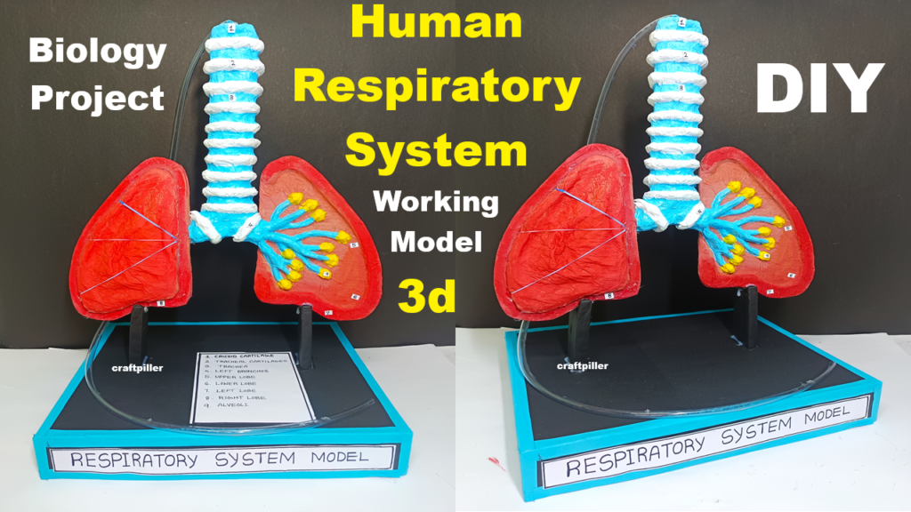
This Human Respiratory System Working Model (3D Lungs) is a DIY biology project made using simple and low-cost materials such as cardboard, aluminium foil, tissue paper, paint, and paint brushes. The model visually demonstrates the structure and working of lungs, making it ideal for science exhibitions and classroom learning.
Aim of the Project
The main objectives of this project are:
- To understand the structure of the human respiratory system
- To explain the breathing mechanism (inhalation and exhalation)
- To create a 3D working model using eco-friendly materials
- To promote hands-on learning in biology
- To improve presentation and explanation skills
Materials Used
The following materials are used to make this model:
- Cardboard (base and structure)
- Aluminium foil (airways and lung texture)
- Tissue paper (lung expansion effect)
- Paint (red, blue, pink, black, white)
- Paint brushes
- Glue and tape
- Scissors and cutter
- Straws or pipes (trachea and bronchi)
- Balloons (optional, for working demonstration)
Overview of the Human Respiratory System
The respiratory system consists of the following main parts:
- Nose and nasal cavity
- Trachea (windpipe)
- Bronchi
- Lungs
- Alveoli
- Diaphragm
The main function of this system is gas exchange, where oxygen enters the blood and carbon dioxide is removed.
Description of the 3D Working Model
The model is mounted on a strong cardboard base. The outline of the human chest cavity is drawn on cardboard. Inside this structure, the lungs and air passages are constructed in a 3D form using aluminium foil and tissue paper, giving a realistic appearance.
Each part of the respiratory system is clearly labeled for easy understanding.
Structure and Function of Each Part
1. Nose and Nasal Cavity
The nose is the entry point of air. It filters dust particles, warms the air, and adds moisture. In the model, the nasal passage is shown at the top using cardboard and colored paper.
2. Trachea (Windpipe)
The trachea is represented using a straw or rolled aluminium foil tube. It carries air from the nose to the lungs. Rings are drawn to represent cartilage rings that keep the trachea open.
3. Bronchi
The trachea divides into two bronchi, one leading to each lung. In the model, this branching is clearly shown using foil-covered tubes.
4. Lungs (3D Structure)
The lungs are the main organs of respiration. In this model, lungs are made using tissue paper layered over aluminium foil to create a soft, expandable texture. Pink or light red paint is used to give a realistic look.
The 3D design helps viewers understand the size, shape, and position of lungs inside the chest cavity.
5. Alveoli
Alveoli are tiny air sacs inside the lungs where gas exchange takes place. Small sponge pieces or clustered tissue paper bubbles are used to represent alveoli. Labels explain that oxygen enters blood here and carbon dioxide leaves the blood.
6. Diaphragm
The diaphragm is a dome-shaped muscle located below the lungs. In this model, it is shown using curved cardboard or stretched tissue paper. It plays a key role in breathing movements.
Working of the Respiratory System
Inhalation (Breathing In)
- The diaphragm contracts and moves downward
- Chest cavity expands
- Air pressure inside lungs decreases
- Air enters the lungs
In the model, lung expansion is demonstrated by gently pulling the diaphragm part or inflating balloons (if used).
Exhalation (Breathing Out)
- The diaphragm relaxes and moves upward
- Chest cavity becomes smaller
- Air pressure inside lungs increases
- Air moves out of the lungs
This working mechanism clearly shows how breathing occurs.
Educational Importance of the Model
- Makes abstract biological processes easy to understand
- Enhances visual and practical learning
- Suitable for middle and high school students
- Improves interest in biology and human anatomy
Advantages of Using Cardboard and Recycled Materials
- Low-cost and easily available
- Eco-friendly and recyclable
- Safe for students
- Encourages creativity and innovation
Applications of Respiratory System Knowledge
- Understanding breathing and health
- Awareness about lung diseases like asthma and bronchitis
- Importance of clean air and pollution control
- Promotes healthy lifestyle habits
Conclusion
This Human Respiratory System Working Model (3D Lungs) effectively demonstrates the structure and working of the lungs using simple DIY materials. By using cardboard, aluminium foil, tissue paper, and paint, the model becomes both educational and visually attractive.
The project helps students understand how breathing occurs and why the respiratory system is essential for life. It also delivers an important message: Healthy lungs need clean air. This model is ideal for biology projects, science exhibitions, and classroom demonstrations.