Let’s explain the workings of the heart in biology terms:
- Heart Chambers:
- The heart is a muscular organ with four chambers: two atria (upper chambers) and two ventricles (lower chambers).
- Blood Circulation:
- Deoxygenated blood, low in oxygen and high in carbon dioxide, enters the right atrium from the body through veins.
- The right atrium contracts, sending the blood through the tricuspid valve into the right ventricle.
- Pulmonary Circulation:
- The right ventricle contracts, pumping the deoxygenated blood through the pulmonary valve into the pulmonary artery.
- This artery carries blood to the lungs, where carbon dioxide is exchanged for oxygen.
- Oxygenated Blood Return:
- Oxygenated blood returns to the heart from the lungs via the pulmonary veins into the left atrium.
- Systemic Circulation:
- The left atrium contracts, sending the oxygenated blood through the bicuspid (mitral) valve into the left ventricle.
- Body Distribution:
- The left ventricle contracts, pumping the oxygenated blood through the aortic valve into the aorta.
- The aorta branches into arteries, distributing oxygen-rich blood to all parts of the body.
- Valves Regulation:
- Valves, including the tricuspid and bicuspid valves, ensure one-way blood flow, preventing backflow between chambers.
- Cardiac Cycle:
- The series of events, known as the cardiac cycle, involves systole (contraction) and diastole (relaxation) phases of the heart chambers.
- Autonomic Control:
- The heart rate and force of contractions are regulated by the autonomic nervous system, specifically the sympathetic and parasympathetic branches.
- Blood Pressure:
- The heart’s pumping action contributes to blood pressure, necessary for the efficient circulation of blood throughout the circulatory system.
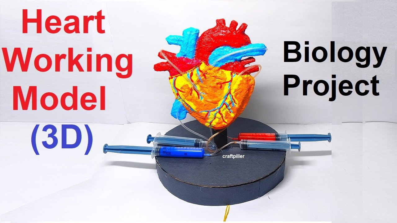
How to make 3d Working model of Heart
Creating a 3D working model of the heart using cardboard and syringes to represent blood pumping involves simulating the pumping action of the heart and the flow of blood through the circulatory system.
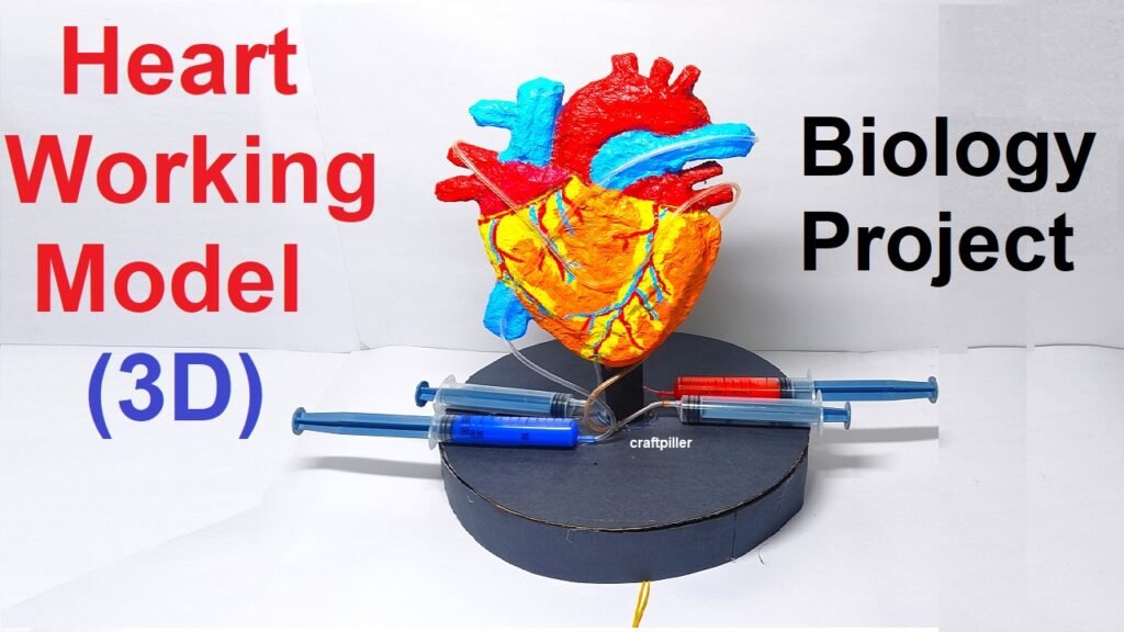
Below are step-by-step instructions for building a simple yet effective heart model:
Materials Needed:
- Cardboard
- Syringes (two or more)
- Flexible tubing or straws
- Red-colored water or liquid
- Clear plastic sheet or plastic bag
- Scissors
- Tape
- Marker
- Glue
- Ruler
- Pencil
Heart Working Model Construction:
- Create the Base:
- Cut out a heart shape from cardboard to serve as the base of your model. This will represent the anatomical shape of the heart.
- Build the Chambers:
- Cut out additional cardboard pieces to represent the atria and ventricles of the heart. Attach them to the base to create the 3D structure of the heart.
- Add the Atria and Ventricles:
- Use smaller pieces of cardboard to represent the atria (upper chambers) and ventricles (lower chambers). Attach them appropriately on the heart base using glue.
- Integrate Syringes:
- Attach syringes to the atria and ventricles using flexible tubing or straws. The syringes will act as the pump for the model. Make sure the syringes are securely connected to the cardboard.
- Create Valves:
- Use additional cardboard pieces to create valves between the atria and ventricles. These valves should open and close to simulate the flow of blood.
- Simulate Blood Flow:
- Connect the syringes with tubing or straws to simulate blood vessels. These vessels should extend from the ventricles to the rest of the model.
- Color the Blood:
- Fill the syringes with red-colored water or liquid to represent oxygenated blood. Ensure that the liquid can be easily seen as it flows through the tubing.
- Cover with Plastic Sheet:
- Cover the entire model with a clear plastic sheet or plastic bag to contain the liquid and prevent any spills.
Model Operation:
- Pumping Action:
- Demonstrate the pumping action by pressing the syringes to simulate the contraction and relaxation of the heart’s chambers.
- Blood Flow:
- Observe how the red-colored liquid flows through the tubing, representing the movement of blood through the heart and circulatory system.
- Valve Function:
- Emphasize the opening and closing of the valves as the syringes are pressed and released, maintaining a unidirectional flow of blood.
This 3D working model effectively demonstrates the basic mechanics of the heart’s pumping action and the flow of blood through the circulatory system. It is a hands-on and visual way to engage learners in understanding the anatomy and function of the heart.
In essence, the heart functions as a dual pump system, facilitating the circulation of oxygenated and deoxygenated blood through distinct pulmonary and systemic pathways, ensuring a continuous and regulated supply of oxygen and nutrients to body tissues and organs.